Methodology
Innovation first
INSERM together with Metatissue, Beonchip and CNRS, have recently developed an original organoid model system, coupled with a microfluidic system enabling the modulation of the organoid by external factors, and the follow-up of its reaction to this external modulation. This innovative technology is based on mesoscale organoids derived from pathological and healthy tissues from patients (Patients Derived Living Material, PDLM) combining vascularization and immune cells focused on the gut tissue.

PDLM
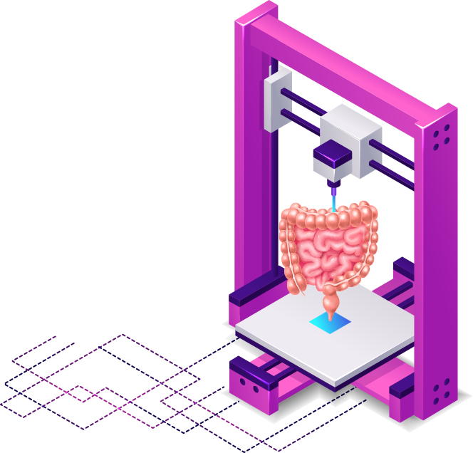
These PDLM will be obtained using an innovative bioprinting method, for large organoids manufacturing (>3 mm), combining multiple small organoids (400 µm) using the S-PIKE technology (Cyfuse Biomedical KK, Japan). This innovative bio manufacturing method, which allows the creation of 3D constructs without scaffolding using only cellular organoids, has already been used to produce several tissues such as trachea, œsophagus, peripheral nerves or blood vessels, but has never been used to produce complex 3D structures such as the gut and its environment.
3D bio-printers
3D bio-printers use bio-inks (alginate, gelatin, collagen, elastin, etc.) to reconstruct a 3D structure with cells. These bio-inks make it possible to mimic the cellular environment and promote cell survival, growth and adhesion. However, they may also have some drawbacks, such as stability and durability for long culture times, nonspecific interactions with cells, potential potency for some, batch variability, and high cost. The S-PIKE 3D bioprinter from Cyfuse Biomedical KK does not use bio-ink, it is the fusion of the organoids between them via the secretion by the cells of their own matrix which leads to the reconstruction of a 3D structure. This technology therefore allows us to overcome the disadvantages associated with bio-inks and to be as close as possible to in situ conditions, by using the patient’s cells secreting their own matrix with the characteristics specific to each individual.
Based on the limitations to develop immunotherapy approaches for human diseases using animal models, we opted for the development of an ex vivo human preclinical model that will reconstitute the physiological complexity of the human gut.
So far, ex vivo epithelial models have been developed from patients and the protocols are well established. P1a-INSERM already mastered ex vivo explant cultures of CD intestinal mucosa. P1c-INSERM has also acquired the expertise in generating gut epithelial organoids from patients or animal models and using them to test innovative therapies. However, these ex vivo epithelial models are not sufficient to test immunotherapy efficacy requiring the presence of immune structures and key tissues that modulate their activity, as well as vascularization.The consolidation of the structure will lead to the formation of a well-organized tissue.
The optimized intestinal organoids (IOs) and PDLM will be integrated into a micro and meso fluidic device permitting vascularization and maintained perfusion, for drug testing.
Recently the development of microphysiological devices has been accelerated and today several industries are marketing this type of system to establish functional vascularization (Mimetas, Emulate, AIM biotech).
The physiological fluidic device designed, is innovative in several aspects since it will circumvent device limitations and allow the addition of an immune/neural environment
Indeed, this device consists of open central chambers, allowing the integration of IOs and PDLMs within their matrix, supplied by micro-channels and meso-channels on each side feeding the chambers to mimic blood microcirculation. The central chambers are equipped with a plug that can be closed tightly. The perfusion will be performed with a microfluidic flow control system. In addition, the device has two chambers connected to each other, one for vascularization of the PDLM, the other to recreate the immune environment.Connected but distinct, they are perfectly adapted for immunotherapy protocol testing. The gut tissues will be obtained from biobanks of patients’ samples biopsies/surgical resections collected by P4-IHU Strasbourg who has already gathered all legal and ethical committees’ approvals.

Project overview
STEP 1
GMP-compliant purification + Characterization and amplification of DP8α regulatory T cells (Tregs) in healthy donors versus IBD patients
STEP 2
Setup and characterization of a relevant ex vivo and animal-free preclinical model for IBD, first as microscopic organoids
STEP 3
Obtention of a macroscopic (mesoscale) 3-dimensional and vascularized organoid structures from the aggregation of the micro-organoids
STEP 4
Production of the first meso/microfluidic device a large vascularized system perfusion, integrating small organoids and mesoscale organoids
STEP 5
Characterization of IBD complex organoid models response to reference therapies and comparison to the IOs and PDLMs preclinical models
STEP 6
Preclinical proof of concept establishment for the efficiency of Treg immunotherapy using the complex gut organoids
